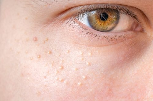
If You Have These Tiny, White Bumps on Your Face, Don’t Try to Remove Them!
You may have experienced milia as an infant, but are unaware of it. A milium cyst, or milia, is generally caused by entrapped keratin (the protein that makes up hair, skin, and nails). It is most common in infants, with up to half of all infants developing it[1]. This is because at this early stage in development the infant’s skin is still learning how to exfoliate. However, milia can occur in people of all ages when something clogs the ducts leading to the skin’s surface, like an injury or a burn[2].
Milia is often seen as tiny white bumps on the nose, chin, or cheeks, and can also be seen on other areas of the body. Though milia are seen in both infants and adults, the types of milia vary, and treatment is different for each and is very often not necessary. Milia is generally completely harmless and will go away on its own. That being said, it’s important to understand how to identify these little bumps to understand if you should intervene or not.
The Different Types of Milia

Milia types are classified based on the age at which the cyst forms or what’s causing the cyst to develop[1]
Neonatal Milia

Neonatal milia develop within infants and clear up within a few weeks. Cysts are typically seen on the face, scalp, and upper torso[2]. According to the Seattle Children’s Hospital, milia occurs in around 40% of newborn babies[3].
Juvenile Milia

Rare genetic disorders, such as Nevoid basal cell carcinoma syndrome (NBCCS), Pachyonychia congenita, Gardner’s syndrome, or Bazex-Dupré-Christol syndrome can lead to juvenile milia[2].
Milia en Plaque

This type of milia is often associated with genetic or autoimmune skin disorders, such as discoid lupus or lichen planus, and it affects the eyelids, ears, cheek, or jaw. It is commonly seen in middle-aged females, but it can be seen at any age in either gender[1].
Primary Milia

This type of milia is seen in older children and adults. Cysts can be found around the eyelids, forehead, or on the genitalia. It may disappear after a few weeks, or last for several months[1].
Traumatic Milia

Milia can sometimes occur on the skin where another injury (such as a rash or a sunburn) have occurred. The cysts may become irritated, making them red along the edges and white in the center[1].
Diagnosis

Due to the fact that milia are quite visible, a dermatologist will visually determine if you have milia based on the appearance of cysts. Skin lesion biopsies are only needed in rare cases[2]. If you see similar little white bumps on your skin you may want to check with a doctor to deduce if you indeed have milia, and to decide on a treatment plan (if you desire one).
Milia Removal and Treatment

Due to the fact that infant milia generally disappear on their own within a few weeks, there is no milia removal or treatment process.
Milia in older children and adults disappears on its own as well, but some people may choose to treat it if there is some discomfort. Common practices include:
- Cryotherapy – Liquid nitrogen freezes the milia. It’s the most frequently used removal method.
- Deroofing – A sterile needle picks out the contents of the cyst. This method is common for treating milia.
- Topical retinoids – These vitamin A-containing creams help exfoliate your skin.
- Chemical peels – Chemical peels cause the first layer of skin to peel off, unearthing new skin.
- Laser ablation – A small laser focuses on the affected areas to remove the cysts.
- Diathermy – Extreme heat destroys the cysts.
- Destruction of curettage – The cysts are surgically scraped and cauterized.[2]
- Milia has even been treated using a paper clip, but it is recommended that the procedure be completed by a doctor and not attempted at home[5]
The following video shows a dermatologist removing multiple milia using the deroofing method.